Michael Wiley, a Surgeon’s Anatomist, Retires
When Mike Willey joined the Division of Anatomy
39 years ago in 1976, Anatomy was a separate department
in the Faculty of Medicine. Shortly afterward, the
amount of time available to Anatomy in the curriculum
was decreased, leading to staff reductions, and administrative
mergers began.
Under Dean Arnie Aberman, the anatomists talked to
Laboratory Medicine, Pathobiology, Medical Biophysics,
Physiology and Surgery about merger. John Wedge was
particularly helpful and facilitated the adoption of the
Division of Anatomy into the Department of Surgery.
“Our Division of Anatomy is the best teaching
Division of Anatomy in the world. The Division of
Anatomy has always had equal standing with the other
divisions in the Department of Surgery with a seat on
the Senior Advisory Committee and the Promotions
Committee. We have a regular rotation in the Grand
Rounds cycle with the Harland Smith Lecture, and our
own budgetary independence. When the University cut
the support of both the clinical and the basic scientists,
the clinical cut was considerably more severe. John
Wedge made sure that the Division of Anatomy was
treated on an equal footing with the basic science departments
rather than the clinical departments.
“We teach dissection with very good laboratory demonstrators.
Occasionally surgical alumni step in to help,
Bob Filler and Alan Hudson among them. Recently,
we’ve had a general surgical resident, Chethan Sathya,
teaching. They have all done a superb job”
Q: You have been widely praised for your teaching
methods. Can you tell us a little bit about them?
A: “The first year medical students focus is the whole
organism. We realize that they won’t all go into anatomically
focused specialties such as Surgery, Radiology and
Pathology. The lectures follow the tradition of Harry
Whitaker, a revered past teacher, who said: ‘Keep it
simple, accessible and logical in its sequence’. I teach
most of the course, so I know what the students have
had and what they will have. The flow is steady, unlike
courses with multiple teachers who repeat and overload
students because their goal is to be sure they get enough
background . We use a well-illustrated note package that
is popular with the students.
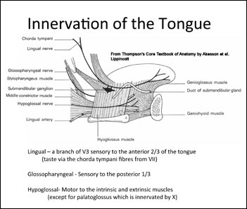
sample of one of the illustrations from the anatomy notes: Innervation of
the tongue
“The virtual microscope is our alternative to the hours
of the old punitive exercise of looking down microscope
barrels trying to focus bad images, without knowing
how to set up the scope, and asking teachers who stagger
between students to identify structures. I found a
company that was working on high resolution pictures
of pathology and other images to be placed digitally into
the medical record. I talked to the company, got a grant
from the University Provost and worked with the company
to modify their program, so that now students do
their histology at home with annotated slides which they
can enlarge and move easily with their computer.
|
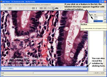
screen shot from the digital histology program that is used for instructing
the students on how to use the program for the first time: duodenum
There is a much lower stress level and they perform
very well on tests. My favorite place to teach is in the
dissecting room, with small groups. The students appreciate
the dissection experience and it gives them the
opportunity to teach each other. This is a very productive
opportunity for exchange of information on more
than just anatomy.”
Q: What are the values that drive the teaching
program?
A: “From the very start, the people in the Anatomy
Department were committed to teaching and very
dedicated to students. The students instituted a Harry
Whitaker Award for the Best Teacher of Anatomy. ”
Mike has won this several times. He is surrounded by
dedicated teachers. The Anatomy Division ranks at the
top of University groups in teaching awards, having won
the Aikins Award, the highest teaching award in the
Faculty of Medicine, a total of 9 times.
The Division has a proud history of publications,
‘Grant’s Atlas’, the reference standard for Anatomy
throughout the world is a product of our anatomists.
It is currently edited by Anne Agur. The specimens
from which the drawings were taken are now in the
JCB Grant Anatomy Museum. The Grant’s Anatomy
artists eventually became the Division of Biomedical
Communications (http://www.surgicalspotlight.ca/Shared/PDF/Summer07.pdf page 7). There is currently
an explosion of anatomy atlases, but Grant’s remains the
premier example. It is not simply an information dump,
but has a very strong teaching component.”
Michael retired on June 30th, 2015, but continues
to teach part-time. His wife is a retired school teacher
with a Master’s Degree in special education. After raising
5 children, when she returned to the classroom, she
developed an expertise in teaching children with autism.
Their 5 children include Joseph, a fellow in Pediatrics at
the Hospital for Sick Children; Daniel who is doing a
doctorate in Communications at New York University;
David, a civil engineer; Kate, a highschool teacher with
a focus on teaching children with autism; and Margaret,
the Residential Program Manager at Camp Oochigeas,
a camp for children with cancer on Lake Rosseau that
runs activities all year, including winter camps , and has
approximately 400 adult volunteer counselors, including
three of her siblings.
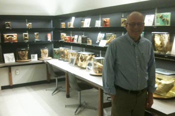
Mike Wiley in the Anatomy museum
Q: What are you currently reading?
A: “Valor Road” by John Nadler, the story of three World
War I Victoria Cross winners from Winnipeg and “Lines
on the Water” by David Richards, a book about fly fishing
and the author’s life growing up in the Miramichi
River Valley, one of Michael’s hobbies. He also plays
old-timers hockey and is developing a vacation property
in Madawaska.
Q: Of your many accomplishments, what is your
proudest?
A: My family. They are all productive contributors to our
community.
M.M.
|